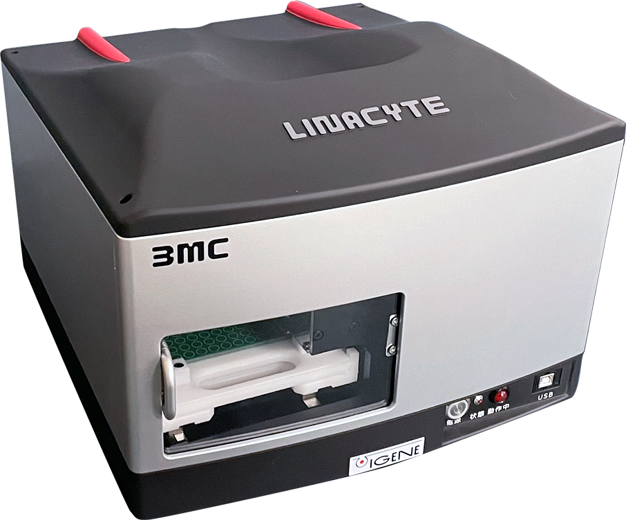
Microplasma Gene/molecular Delivery System LINACYTE 3M

Plasma-Driven Molecular Delivery Mechanism of Linacyte 3MC

Features

A Unique Mechanism of Gene Uptake by Cells
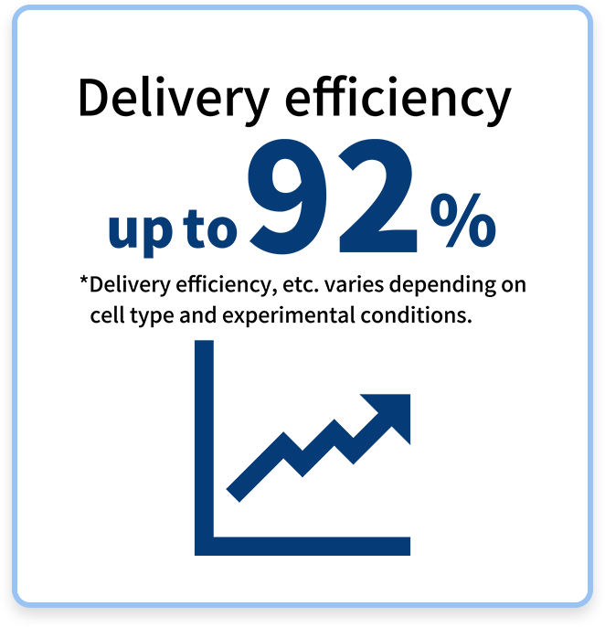
01High efficiency

LINACYTE 3MC achieves both a high success rate for transfection and cell viability.
In conventional methods, there was a problem of increased cell damage when trying to increase delivery efficiency, but this product's unique technology enables highly efficient delivery while maintaining the viable cell rate.
If the viability rate is maintained at 80% or more, the delivery efficiency is 50% or more, and depending on the cell, it is possible to deliver even higher rates.

Delivery efficiency up to 92%

02Safety

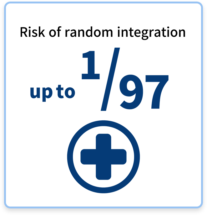
Microplasma technology can significantly reduce the risk of random integration compared to conventional electroporation and lipofection.
The brightness after 25 days of GFP expression is extremely low, about 1/97 that of electroporation and about 1/41 that of lipofection, significantly improving safety. This characteristic minimizes damage to cells and tissues, ensures efficient gene delivery, and increases the accuracy and reliability of research.

Risk of random integration is up to 1/97

03Cost-effectiveness

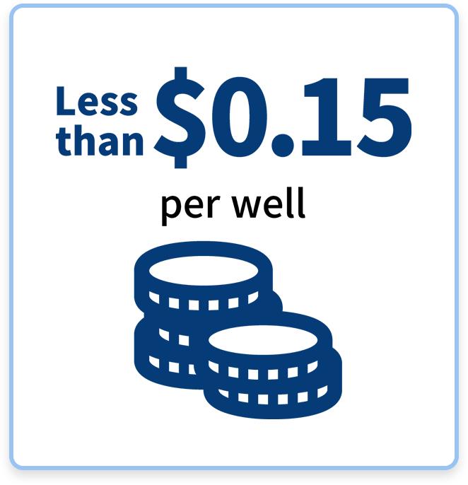
LINACYTE 3MC achieves low running costs because it does not require special equipment or expensive reagents.
The cost per well is Less than $0.15, making it very economical and easy to introduce in existing research environments.
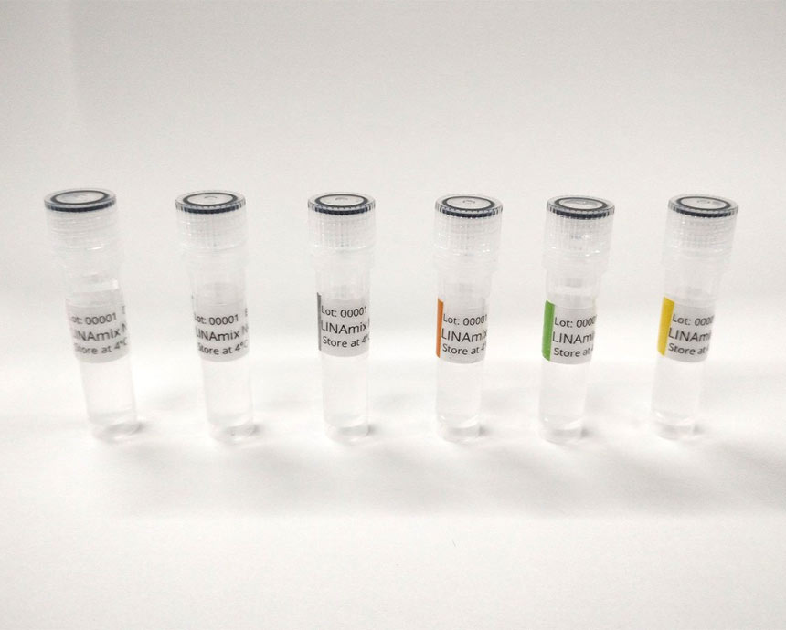
The only required reagent we provide is a buffer mixed with the plasmid or target molecules.

Less than $1 per well

04High-speed processing

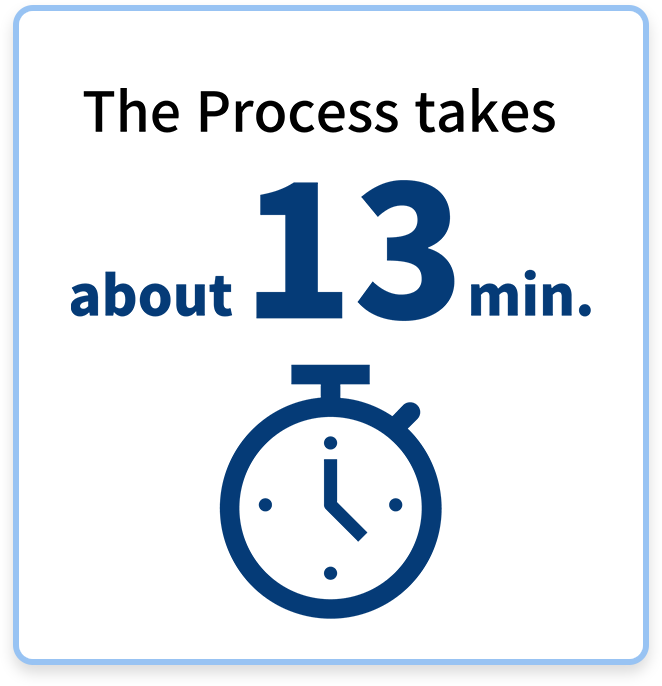
With LINACYTE 3MC, the process from plasmid addition to the end of the delivery process takes only about 13 minutes per 96-well plate.
This short processing time makes it possible to efficiently process an entire 96-well plate. For researchers, this short time to complete the work contributes to improving experimental productivity, and results can be obtained quickly, especially when dealing with large amounts of samples. A major advantage of this method is that it significantly saves time and effort compared to conventional methods.

The Process takes about 13 minutes
Comparison with conventional methods
plasma Commonly used gene introduction methods
Viral vector Lipofection Electroporation
Efficiency High High High~Low High~Low
Cytotoxicity Low Low Mid High
Operability Easy Standard Easy Easy
Side effect Low Mid Unknow Mid
Cell type Over 70 types of cells. Include blood cells and primary cultured cells. Restricted by virus Low efficiency with blood cells and primary cultured cells Low efficiency with blood cells and primary cultured cells
etc Complet in a short time P2 level or higher equipment required High cytotoxicity, expensive reagents Large amount of cells required, expensive reagents and equipment
List of Transfected Cells

Yellow: epithelium Blue: floating Red: fibroblast Green: neuroblast Purple: primary, stem cells, others

No. Cell name Origin Tissue of origin
1 HepG2 Human liver
2 HuH7 Human liver
3 FLS3 Mouse liver(fetus)
4 L alpha Mouse connective tissue
5 MG-63 Human bone
6 Hela Human uterus
7 MDCK Dog kidney
8 HEK293 Human kidney(fetus)
9 HSC-3 Human tongue
10 SAS Human tongue
11 CaCo-2 Human colon
12 A375 Human skin
13 G361 Human skin
14 HSC-5 Human skin
15 CHO-K1 Hamster ovary
16 DD762 Mouse mammary gland
17 HCC1937 Human mammary gland
18 PANC-1 Human pancreas
19 T24 Human bladder
20 BJAB Human peripheral blood
21 Jurkat Human peripheral blood
22 K562 Human peripheral blood
23 MOLT-4 Human peripheral blood
24 THP-1 Human monocyte
25 RAW Human monocyte
26 U937 Human monocyte
27 TK6 Human blood cell, lymphoid system
28 EOL-1 Human eosinophil
No. Cell name Origin Tissue of origin
29 L6 Rat Muscle tissue
30 L Mouse connective tissue
31 L-929 Mouse connective tissue
32 COS-7 Green monkey kidney
33 3T3L1 Mouse fetus
34 NIH 3T3 Mouse fetus
35 Lu99A Human lung
36 SF-TY Human skin
37 Mewo Human dermal lymph node
38 MC3T3-E1 Mouse skull
39 COLO201 Human colon
40 Hu09 Human bone
41 MBT2 Human bone
42 PC-12 Rat adrenal gland
43 GOTO Human nerve
44 SH SY5Y Human nerve
45 Fibroblast Human skin
46 HDF Human skin
47 NHEM Human melanocyte
48 Huvec Human endothelial cell
49 Tic Human iPS cells
50 ACS Human Adipose stem cells
51 HaCaT Human skin keratinocytes
52 Astrocytes Human iPS cells
53 Neurons Human iPS cells
54 Nerve cell Rat nerves
55 MS5 Mouse bone marrow stromal cells
       
Product specifications
LINACYTE 3MC
Dimensions 360mm (D) x 340mm (W) x 260mm (H)
Weight ~9.8kg
Japan 100V, 50/60Hz 11W
Overseas 100-240V, 50/60Hz 11W
Plates 96-well MTP
Cells Animal cells(Include blood cells and primary cultured cells.), etc.
Number of cells / A process 3 million cells/plate (depending on cell type)
Patent Patent No. 6189019
Accessories
Gene delivery buffer kit LINAmix AD Buffer OptKit
6 types for adherent cells (LINAmix N-kp / N-xz / N-jf / N-ql /N-ry / N-mt Buffer)
LINAmix SU Buffer OptKit
6 types for suspension cells (LINAmix N-ry / N-mt / N-vd / N-bg / N-nc / N-hw Buffer)
Gene delivery buffer LINAmix N-kp Buffer
LINAmix N-xz Buffer
LINAmix N-jf Buffer
LINAmix N-ql Buffer
LINAmix N-ry Buffer
LINAmix N-mt Buffer
LINAmix N-vd Buffer
LINAmix N-bg Buffer
LINAmix N-nc Buffer
LINAmix N-hw Buffer
Plates We recommend 96-well plates. Please contact us for details.

*We can also provide custom-made buffers.

Frequently Asked Questions
What is a microplasma gene/molecular delivery device?
The microplasma gene/molecular delivery device is a technology that uses microplasma discharge to delivery genes and molecules into cells. Its special feature is that it allows rapid and efficient gene delivery without the need for special reagents or viruses.
Why do you use microplasma?
It has been confirmed that microplasma induces endocytosis.
This allows for the uptake of molecules closer to nature.
What are the main advantages of this device?
The main advantages are as follows:
High gene delivery efficiency
High safety
High cost performance
High speed processing
What is the difference compared to other gene delivery methods (electroporation, lipofection, etc.)?
Compared to other methods, this device has low cost, short time, high cell viability, and wide applicability.
Since it does not require viral vectors or chemical reagents, it increases the flexibility of experiments.
What types of cells can be used?
It can be used with adherent cells such as HEK cells, L929 cells, and CHO cells, as well as suspension cells and primary cells. Please contact us for details.
Is there any damage to cells?
Cytotoxicity is low, and cell viability is higher than that of conventional methods.
However, since it depends on the type of cell and conditions, please check the details before the experiment.
Do I need special reagents or consumables?
No, no special reagents or expensive consumables are required. Regular culture plates can be used as is.
How long does the gene delivery process take?
One process takes about 13 minutes per plate.
Is it difficult to operate the device?
No, the operation is very simple and does not require special skills or experience. Basic operating procedures are described in the manual.
Does the device require maintenance?
Normally, regular cleaning and inspection are sufficient.
Can I try it out before purchasing?
Yes, we have a demonstration device available. Please contact us for details.
I would like to know the price and introduction cost of the device.
The price of the device itself is 2,980,000 yen (3,278,000 yen including tax), and one bottle of dedicated reagent (enough for five 96-well plates) is 8,000 yen (8,800 yen including tax).
If you would like a quote, please contact us using the inquiry form.